December 2025 · Bioinformatics & Comp Bio· Education & Pedagogy· Interactive Simulation· Web Development
Interactive Viscerosomatic Atlas
Bridging Clinical Microbiology and Osteopathic Manipulative Medicine (OMM)
Interactive Viscerosomatic Atlas (Posterior Integration)
Summary
The Interactive Viscerosomatic Atlas, part of the "NEURO-FORGE" suite, is a specialized clinical learning tool designed to bridge Clinical Microbiology and Osteopathic Manipulative Medicine (OMM). Featuring a dual-view (Anterior/Posterior) reactive SVG interface, it simulates clinical palpation by asking students to correlate specific microbial pathogens (e.g., Strep pneumoniae, E. coli) with their corresponding viscerosomatic reflexes and Chapman points. Through a gamified "Hot/Cold" feedback system and bilateral associative logic, this interactive platform reinforces critical pattern recognition, translating abstract anatomical relationships into tangible digital muscle memory.
1. Executive Summary
This feature is a specialized interactive learning tool designed to bridge the gap between Clinical Microbiology and Osteopathic Manipulative Medicine (OMM). Unlike standard static anatomical charts, this tool utilizes a “Hot/Cold” reactive SVG interface to simulate clinical palpation and diagnostic reasoning, forcing students to correlate specific pathogens with their corresponding somatic dysfunctions.
NEURO-FORGE
Clinical Vignette
35M with frontal headache. Identify sympathetic levels for Head & Neck.
Targets Found 0 / 5
2. Unique Value Proposition
- Reactive “Hot/Cold” Feedback: The system provides immediate visual feedback during the search phase. Hovering over a correct anatomical zone triggers a Neon Yellow cue (positive likelihood), while incorrect zones trigger a Neon Red cue (negative/warning). This mimics the clinical process of “ruling in/ruling out” diagnoses based on palpation.
- Bilateral Associative Logic: Clicking a unilateral structure (e.g., Left Kidney Chapman point) automatically activates the contralateral side. This reinforces the physiological reality that viscerosomatic reflexes often present bilaterally.
- Cross-Disciplinary Integration: This is likely the only web-based tool that directly links specific microbial agents (e.g., Streptococcus pneumoniae) to specific muscular hypertonicity patterns (e.g., T2-T7 Ropy Paraspinals), integrating two distinct medical board subjects into one clinical vignette.
3. Operational Guide
- Select Pathogen: The user selects a clinical case (e.g., “E. coli - Pyelonephritis”) from the dropdown.
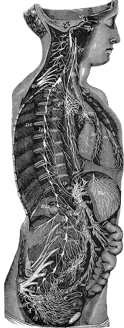
- Select View: The user toggles between Anterior (Nerves) for sympathetic chain involvement or Posterior (Muscle) for somatic dysfunction.
- Scan & Palpate: The user moves the cursor over the vintage anatomical map. The “Hot/Cold” neon system guides them toward the correct level without revealing the answer immediately.
- Confirm Diagnosis: Clicking the target locks the zone in a Persistent Neon Yellow state.
- Clinical Correlation: Once all targets are found, a “Clinical Pearl” card unlocks, explaining the physiological mechanism (e.g., why renal inflammation causes Psoas spasm).
4. Educational Utility in Osteopathic Medicine
- Visualizes the Invisible: Students often struggle to visualize how an internal organ infection translates to back pain. This tool makes that relationship concrete.
- Reinforces Pattern Recognition: By repeating the motor action of clicking T2-T7 for pneumonia or T10-L1 for kidney infection, the student builds “digital muscle memory” that parallels clinical rote memorization.
- Immediate Remediation: The “Red Flash” on wrong clicks provides instant, low-stakes failure, correcting misconceptions (e.g., looking for kidney reflexes in the neck) in real-time.
5. Future Improvements
- 3D Rotation: Replacing the static 2D SVG with a 3D model would allow students to trace the nerve from the anterior ganglia to the posterior spine.
- Haptic Feedback: On mobile devices, adding vibration when hovering over a “Hot” zone would further simulate the sensation of finding a somatic dysfunction.
- Randomized Case Mode: A “Challenge Mode” that hides the disease name and asks the student to diagnose based only on the highlighted muscle pattern.